CEREBRAL BLEED
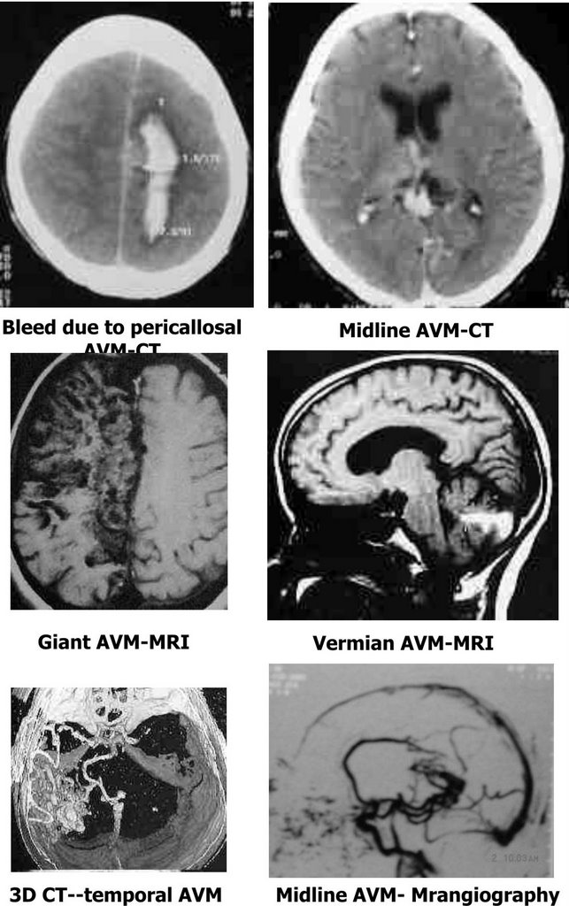
CT scan –may suggest a nidus as a low density within the hematoma (nidus sparing sign). A serpiginous enhancing lesion with an early draining vein; associated hypoperfused areas may be evident as low-density areas.
MRI scan –T1 and T2 images may show areas of flow void; associated hemorrhage including subclincal hemorrhage and areas of cortical atrophy and hypo perfusion are better seen.
MRAngiography and 3D CT may outline the AVM; better suited for follow up studies.
Digital angiography is still the imaging mode of choice. A detailed study of the arterial feeders, the nidus and venous drainage is mandatory.
SPECT and functional PET scanning are useful for assessment of cerebral perfusion.
posted by irratia at 1:21 AM
![]()

0 Comments:
Post a Comment
<< Home